Radiographic Images
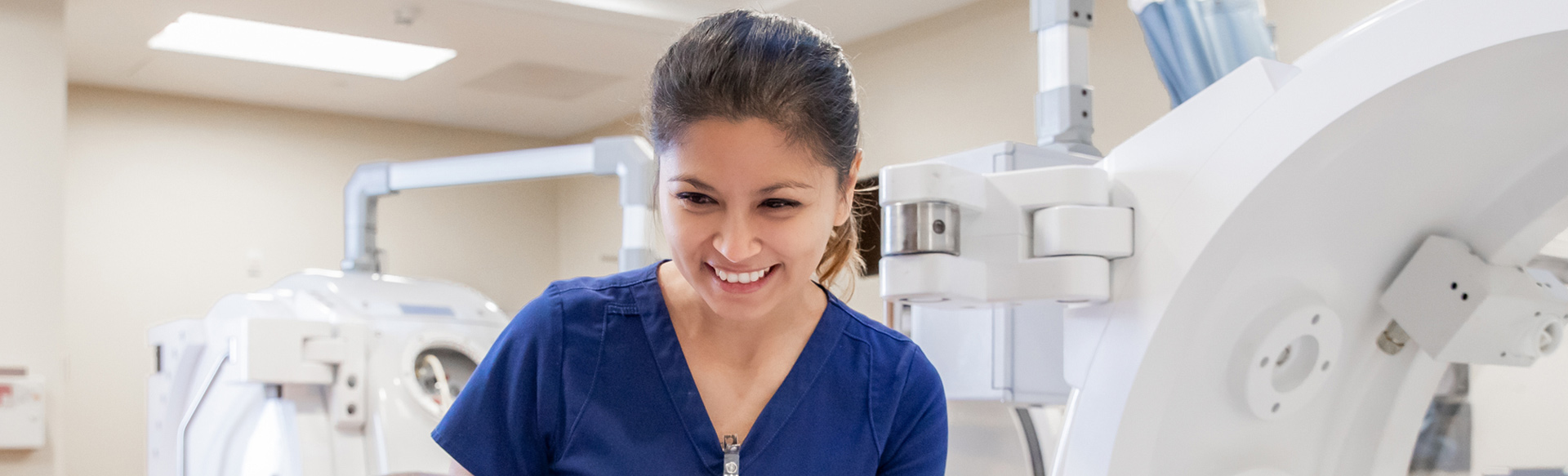
Radiology is an important part of diagnosing and treating your orthopedic
concern. We use several imaging tests to provide us with detailed images
of your condition or injury. With diagnostic radiology, we take images
to identify and diagnose your health concerns. With therapeutic radiology,
we use imaging during treatment or surgery to guide your procedure.
What is an X-ray?
An X-ray is one of the most used radiology exams. X-rays provide a static
image of your bones and organs.
What is a CT scan?
CT stands for computerized tomography. A CT scan takes X-ray images from
several different angles during a single scan. These images are then combined
using a computer to create detailed images of your body.
What is an ultrasound?
An ultrasound is also known as a sonogram. It’s a very common procedure
that provides real-time images of your tissue, blood flow, and organs.
It uses sound waves to produce dynamic images.
What is an MRI?
An MRI, or magnetic resonance imaging, provides images of your body without
using X-rays. Instead of radiation, MRIs use magnets and radio waves to
create images. Radio waves are sent into the body and then back. A computer
takes these returning signals and converts them into images. Your doctor
may use an MRI to create images of almost any part of your body.
What is nuclear medicine?
Nuclear medicine is a field of radiology that uses radionuclides, which
is a specific type of atom that emits radiation. These atoms collect in
specific body tissues.
One of the most common nuclear medicine procedures is a PET scan, which
stands for positron emission tomography. Using a small amount of radioactive
material, PET scans create detailed images of your body’s function.
PET scans are painless and noninvasive.